Incidental finding of a superior lumbar hernia (Grynfeltt-Lesshaft hernia). In this case, only a lobule of retroperitoneal fat is herniating through the defect, but organs can also herniate through.
Continuing the theme of things extending into spaces they don't belong in, this is an incidental finding of an inguinal hernia that contains a small portion of the bladder. The patient got the CT for other reasons.
Bowel into inguinal hernia causing bowel obstruction.
Appendix into inguinal hernia, incidental finding.
Ventriculoperitoneal shunt into inguinal hernia, incidental finding.
[LEFT]: This patient had one of the longer cerebellar tonsillar herniations I've seen. The tonsil is peg-like in shape and extends quite far below the foramen magnum to the level of the C2 posterior arch. As a result, there is crowding at the foramen magnum that is enough to impede CSF flow, resulting in hydrocephalus with dilated ventricles. Partly seen in the cervical cord from C2 and below is a syrinx, an associated finding. Chiari I is thought to be due to not enough space provided for the cerebellum by the calvarium or skull base shape, causing it to herniate into the spinal canal and cause trouble.
[RIGHT]: A comparison normal from online for you to compare the cerebellar tonsils.
[LEFT]: The midbrain has a deep interpeduncular cistern, and the superior cerebellar peduncles are very prominent and elongated, making the brainstem at this level look like a molar tooth. This is a classic finding in Joubert syndrome.
[RIGHT]: A comparison "normal" midbrain. However, this patient's brain is not normal at all. Can you find the abnormalities?
Answer
Compare the left and right temporal lobes in [RIGHT] to the [LEFT] image. Look at how many more gyri and sulci there are in the [LEFT] image. The [RIGHT] patient has a diffuse pachygyria (abnormally reduced brain gyrations). Both Joubert syndrome and pachygyria arise from failure of neurons to migrate, although the genes involved and underlying mechanism are different between the two. (NB: Pachygyria is just a descriptive term for less than normal number of gyri, which can be from a large number of causes mostly having to do with abnormal neuron migration.)
Female in her 30s with painful left shoulder.
[Left]: X-ray shows a mass arising from the left proximal humerus and extending into the adjacent shoulder soft tissues with really aggressive periosteal reaction ("hair on end"). The proximal humerus itself is also heterogeneous with lucent areas. The lateral surface of the upper humerus shows "saucerization," where the cortex is thinned out and looks like a saucer seen on edge.
[Middle]: MRI IR sequence shows a hyperintense bony mass with large soft tissue component.
[Right]: MRI postcontrast T1 IDEAL shows that the mass is enhancing.
This turned out to be high-grade surface osteosarcoma.
33 year old female with abdominal pain, abdominal distention, nausea/vomiting, early satiety, and weight loss.
Bottom right: Ultrasound done in a panorama shows how distended the abdomen is by a large multi-cystic mass.
Top right: Non-panoramic ultrasound image shows how limited the imaging modality is in being able to cover such a large mass. This image also shows a more solid area within the mass.
Left: CT images approximately where the ultrasound was done.
The patient underwent laparotomy with removal of the ovarian, fallopian tube, and appendix. There was a large ovarian cyst that was draining serous fluid (watery), mucinous fuid (mucus-like), and blood. The final path was as titled.
Postcontrast imaging of 2 patients with glioblastoma. These tumors are notorious for spreading along the white matter tracts - in this case the transverse fibers of the corpus callosum, given them a classic "butterfly" appearance.
I think while the general communities have made it, a lot of niche communities failed to attract enough population to keep on generating more content. As an example, just search for the "Imaginary" series of landscape art communities on the Fediverse (eg. ImaginaryVistas). Many of them don't have any recent posts or 1 post per days or weeks. That's not enough to keep people invested. Even the largest digital art community is still mostly carried by 1 person.
The Lord said that the "VCR shall be saved" with the knife technique, but in the following paragraph, it was not the VCR that was saved, but the man that was saved. The VCR was not saved!
Two different patients with genetic disorders resulting in overgrowth of the brain.
These represent mutations in cell cycle and cell metabolism genes that lead to larger cells and/or more cells. These types of disorders tend to have mosaicism of some form, which is to say some cells have the mutation active while others don't. The distribution of these cells can be very geographic/regional - in these two cases, one hemisphere of the brain is involved.
Compare this against a previous case with hemispheric atrophy.
It looks like it could fall over with any gust of wind and kill someone.
Hello everyone!
I am amazed at how quickly this rather specialized community has grown. It gives me perverse pleasure to see that C/Radiology has somehow exceeded C/Medicine in subscriber numbers! So thanks for visiting and allowing me to share my interest in this field with you!
As the community has expanded, we have, of course, come across typical growing pains, and since this is a medical community, some additional factors must also be considered, such as respect for any patient discussions and medical privacy. We have the potential for a lot more growth, but we must be vigilant in respecting medical laws as well. To that end, I have made additional changes to the Community Rules to better clarify the situation for everyone. Additionally, I have conversed with the Lemmy.World admins, who are supportive of this community and now aware of its unique characteristics and requirements.
One major change that has come out of that discussion is that we worry about how inadvertent posts that breach patient confidentiality would behave with federation. It's not like Reddit, where the post is centralized, and there's only one copy to remove. As a consequence, for now, I have changed this community to only allow moderators to post. My hope is that, in the not-too-distant future, Lemmy itself will implement a way for users to post pending moderator approval. Visitors may still comment upon any posts in this Community, and so as a workaround, I've started this megathread for any general questions or discussions you might have regarding radiology. (Please follow the rules still!) If you would like to share a case as a post - please DM me, and I will post on your behalf.
Now onto future updates: For the next few weeks, I will have reduced posting - because I'm going to be away from steady internet. I will continue to post interesting cases I come across thereafter. Eventually, I also plan to have a sticked general guidance on how to look at radiologic images so that you can have a better understanding and capability of looking at these images yourself!
While we're at it, I'm also looking for additional mods to help. I would prefer that you have some medical imaging background, medical background in general, or moderator experience if possible!
Should the instances that responded to you be refederrated? I’m pretty sure I saw some of them on lemmy.world’s block list. I think it would be sad for these small servers to not realize they are, in fact, not connected to the greater fediverse. On the other hand, if you’re an admin, and you don’t know what you’re doing to the point of not knowing your server was infected by hundreds of thousands of bots, maybe it’s too dangerous to refed.
Quick one today. Take a look at Patient A and Patient B.
Patient A has a smooth focal indentation of the posterior cervical esophagus.
Patient B has a broader indentation that is also irregular and nodular along its contour.
Patient A has a cricopharyngeal bar, which is a prominence caused by the cricopharyngeus muscle that can cause dysphagia if it gets really prominent. Patient B has esophageal squamous cell carcinoma.
Patient was a young adult working in finance at a major tech company found to be mute and diaphoretic.
Physical exam notable for fever, tachycardiac, hypertension, awake but not following commands, aphasic, and with hyperreflexia and muscle ridigity. CK peaked to 11,344.
MRI shows multiple ovoid to splotchy confluent lesions in the white matter with diffusion restriction. Lesions also enhanced with hyperperfusion (not shown).
Urine drug test positive for cocaine. Infectious work-up was negative. Steroids were started with good recovery.
Patient denied knowingly taking cocaine but did say weekly use of what they thought was MDMA with friends...
Final diagnosis: Levamisole-induced leukoencephalopathy. Levamisole is an antiparasite medication that is no longer used in the US but still in some other countries. It is a common cutting agent in cocaine. It's neurotoxic effects primarily come from causing demyelination.
I remember this episode quite well because it happened around the time I decided to get into the medical field. In the episode, a young teacher had a first-time seizure while in the middle of teaching. House and team attempted to get a brain MRI, but she got an allergic reaction from the IV contrast. Thereafter, some drama happens, and at some point, they break into her house, find out she's been eating raw pork (wtf?), and diagnose her with the tapeworm infection associated with eating raw pork, cysticercosis (and neurocysticercosis, since it also involved her brain). They took an x-ray of her leg to show all the parasites in the muscles, and then House scolds her for being stupid. I remember thinking that was such as crazy medical story.
The reality is - they could have just repeated the brain MRI minus the contrast part, and the radiologist would have been able to identify neurocysticercosis without issue. House would have complained to Cuddy that she really was wasting his time with these basic cases, and the episode would have lasted 15 minutes tops...
Anyhow, this is a 25 year old Hispanic from jail. Just like the House episode, he presented with first time seizure and headaches.
CT of the head [top] shows a cystic lesion in the left frontal lobe. If one pays attention, one can see a small dot (blue arrow) within the cyst representing the scolex of the tapeworm parasite. Just from the CT appearance, history of seizure, and risk factors of jail (the parasite thrives in areas of low sanitation) and Hispanic (the parasite is endemic to South America), neurocysticercosis is the top possibility. A differential diagnosis of cystic brain tumor is provided to complete the picture.
MR [middle and bottom] shows a cystic lesion again. After giving IV contrast [middle right], one can see the cyst has a thin wall of enhancement (teal arrows). On T2 [bottom left] and especially FLAIR [bottom right], one can see a rim of swollen brain (green arrows) from the inflammation going on around the parasite.
This was diagnosed as neurocysticercosis in the colloidal vesicular stage and antiparasite medication was started.
Oh good to know. I guess I only really caught reddit downtimes in the past.
I like how it looks exactly like reddit's status page, especially considering they just released old.lemmy.world.
[Top]: X-ray shows a lucent, bubbly, lesion of the distal femur at the metaphysis. On the frontal view [top right], there is breakage through the medial femoral cortex into the adjacent soft tissues, not a good sign.
[Bottom]: MRI shows a multicystic lesion filling the distal femur containing multiple locules, many with fluid-fluid, fluid-debris, and fluid-hemorrhage levels. The most common lesions with this striking appearance are aneurysmal bone cyst, giant cell tumor, or telangiectatic osteosarcoma. Unfortunately, there is clearly extension of the bone tumor beyond the bone (yellow arrows), which favors a more aggressive neoplasm from that differential diagnosis - this turned out to be telangiectatic osteosarcoma.
5 year old who fell off a slide.
Initial imaging shows a comminuted fracture through the distal humerus, compatible with a supracondylar fracture. Nothing else appreciable here, except maybe in retrospect some lucency of the distal humerus where the fracture is.
4- and 7-month follow-up radiographs shows a growing lucent lesion of the distal humerus, expanding the bone there. It has a multicystic appearance. A diagnosis of large simple bone cyst versus aneurysmal bone cyst was proposed.
12 month follow-up was done after the cyst was opened surgically, its contents scraped off, and the resulting cavity was packed with allograft bone chips. At surgery, this turned out to be an aneurysmal bone cyst.
5 year follow-up shows involution of the cyst cavity with some residual heterogeneity and a bone spur at the anterior aspect of the distal humerus.
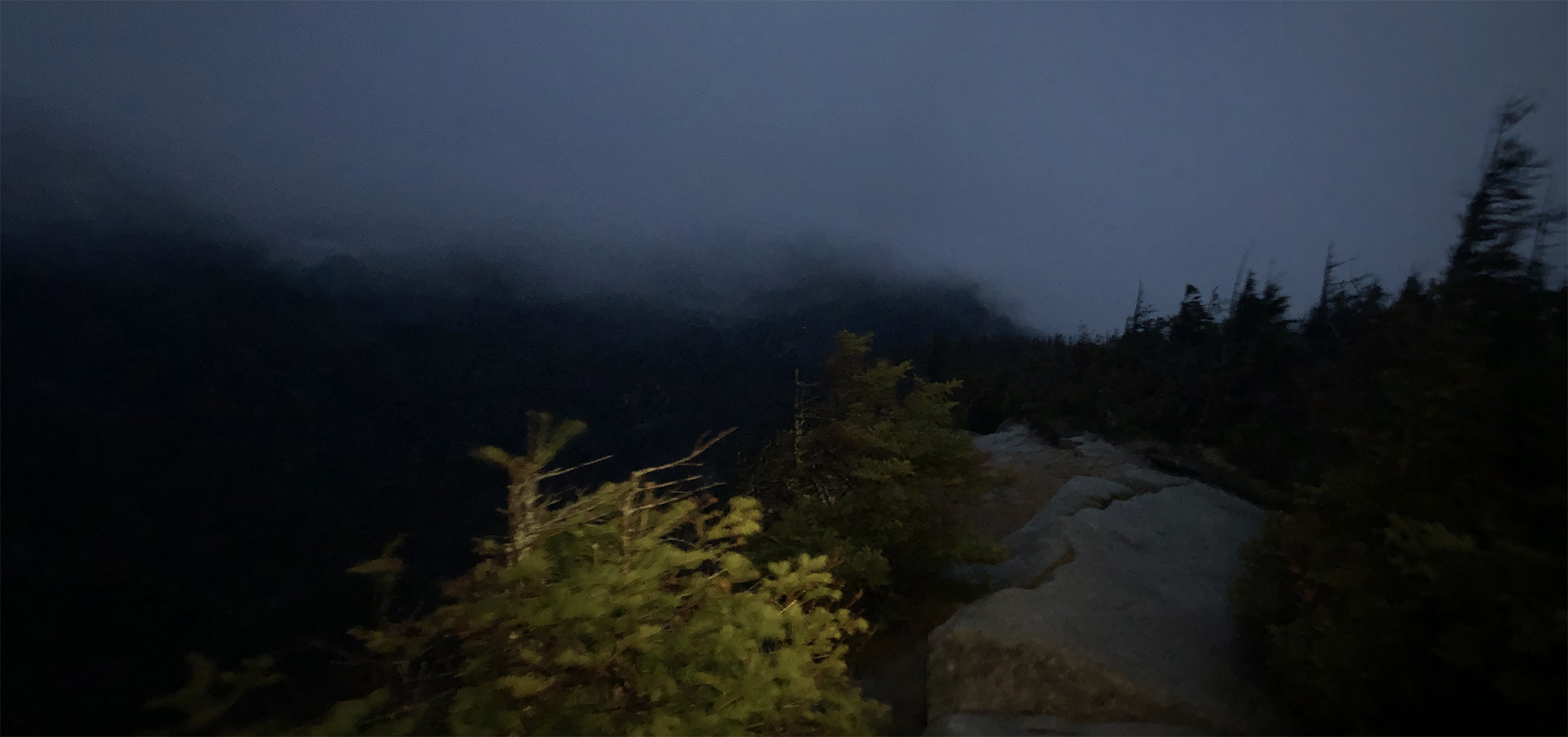
Luckily I did bring my flashlight and batteries, otherwise I would have been screwed. I did not plan to stay up so late, but the ascent to the ridge hike took longer than anticipated, and of course, what was a sunny day in the morning progressed to clouds and rainstorm by the afternoon, which continued into the night.
I continued as best as I can. I was acutely aware of the danger of a trip and fall that might break an ankle and hypothermia - some parts of the hike still had snow, and anytime I stopped, I could feel the cold seeping in.
In the end, I reached my car at around 10 PM.
Getting caught in a large rainstorm in the evening while on a mountain trail with 3 more hours of rocky descent to go.
[Left]: Head CT shows left hemispheric volume loss. The injury happened early enough that even the skull is smaller on that side.
[Right]: Brain MRI shows the severe left hemispheric atrophy. Some of the brain gyri have bulbous ends and a thin neck, resembling mushrooms, a shape called ulegyria and consequence of the brain atrophy. The left lateral ventricle is mildly enlarged due to the atrophied brain.
Red arrows point to 2 big gallstones, top one in the gallbladder and bottom one obstructing a small bowel loop, and a small gallstone in the cystic duct.
Red lines point to hernia entry. Red arrow points to where the bowel tapers and becomes obstructed as it enters the hernia sac.
[Left]: Fetal MRI (FIESTA sequence) shows twins joined from their lower chest to the pelvis, but truly fused and sharing a single abnormal pelvic region. Not shown, but there are 3 lower limbs - one of the twins only had a single lower extremity.
[Right]: Postnatal small bowel follow-through (SBFT). It was unclear initially whether the twins shared a single rectum or had their own rectum. Therefore, contrast was administered via nasogastric tube for the twin with the suspected nonfunctional rectum, and serial imaging was performed until it passed into what turned out to be a separate, functional, but small rectum/anus.
I do not know too much about conjoined twins - not my area of expertise, but the general forms to consider are the side of fusion: ventral (front to front), lateral (side to side), dorsal (back to back), or caudal (tail end to tail end). Within these first 3, there are subtypes depending on how far up the fusion goes (head, chest, abdomen/pelvis); by definition, the caudal version obviously is only a lower body fusion. Once this is derived, an additional classification is the number of upper and lower limbs.
I just experienced my first actual report. 🙃
No clinical history saved on this one - sorry.
[Right] Small bowel follow-through (SBFT), where the patient drinks barium, and then we wait a bit until that barium is in the small bowel, then we take some pictures. This study is showing a long segment of terminal ileum that is strictured and severely narrowed in fibrostenotic Crohn's (red bracket). This is called the "string sign."
[Left] Coronal CT performed sometime after the SBFT. You can still see some residual barium in the small and large bowels (blue arrows). Red bracket shows the CT appearance of the terminal ileum stricture. On the CT, you can also see that the strictured segment has submucosal fat deposition, the "fat halo sign."
I didn't save any clinical history for these - sorry.
[Top] Patient 1 - Gigantic mass along the lesser curvature of the stomach. Look down at your belly - this mass is about 1/3rd the width from left to right.
[Mid] Patient 2 - CTs showing gently lobulated and undulating wall thickening of the gastric cardiac and fundus. Notice the transition from the normal gastric rugae to the smoother wall thickening where it is infiltrated by lymphoma. There is also mild (aneurysmal) dilation of the stomach where the wall thickening is located.
[Bottom] Patient 2 - PET-CT. The wall thickening is ridiculously hypermetabolic with a max SUV of 21.3. For comparison, the liver is normally in the range of 2-4 mean SUV.
Tuberculosis, sarcoidosis, lymphoma, and metastatic disease - these 4 can look like almost anything.
The Voyager app just recently added the ability to block a community directly from the feed without opening the post or the community.
The only person I met with that name, and only in small amounts, is Henrietta Lacks.
I feel like it would be a bot purge or lemmy.world and several other instances being down from the hacking.
Consider undervolting (via Throttlestop or Intel XTU) to prolong your laptop’s longevity and possibly mildly increase its performance. For the same CPU workload, undervolting will reduce the amount of heat generation and therefore the temperature of the CPU, thereby decreasing the risk of hitting the CPU’s temperature throttling and risk of CPU damage.
There are ready guides on youtube and r/gaminglaptops sub, but I’ll leave reddit links out for now. Just search for your laptop model since the exact values will depend on the model and also on luck. If you’re lucky, you can undervolt a lot without causing instabilities.
You don’t see anything from the bot because lemmy.world already defederated from lemmit.online.
-
Do the maneuver @Dadbod89@lemmy.world suggested.
-
Try Flonase. Helps a lot but takes 1-2 weeks to start to work.
-
See a doc if things persist.
That drone live view was pretty nice.
I was really surprised to hear this, so I dug a little deeper. It looks like an honest mistake.
Here's what their admin said:
We unintionally did until about 8 hours ago.
Lemmy.ml has a ton of bot crawlers in its nginx logs spam fetching posts, so I added the bots to an nginx block. Turns out one called kbinbot wasn’t actually a crawling bot, but their federation requester.
If we don’t respond quickly, its because we have notification backlogs that are months long at this point.
Just a week and a half ago, we were celebrating going from #2 to #1 most populous instance. And just 2 weeks before that, I had no idea what a lemmy was.
It's like catching a nice surf wave.
I'll bet you it's because people are lazy and just click that button instead of spending time to type out why they reported.
Source: Definitely accidentally clicked that button when reporting.
